


Apr. 21, 2022
Publisher: Shiseido
R&D/Supply Network
Mariko Egawa, Ph.D., Senior Researcher at Shiseido, receives MEXT Award
Recipient of the Award for Science and Technology in research as a part of the 2022 “Commendation for Science and Technology by the Minister of Education, Culture, Sports, Science and Technology”

Mariko Egawa, Ph.D., a Senior Researcher at the Shiseido MIRAI Technology Institute, received the Award for Science and Technology (Research Category) as a part of the 2022 “Commendation for Science and Technology from the Minister of Education, Culture, Sports, Science and Technology (MEXT)” after being nominated by The Spectroscopical Society of Japan. This award is given to individuals who performed original research and development with a high probability of contributing to the advancement of science and technology in Japan. The recipients of this award were announced on April 8, and an award ceremony was held on April 20 with MEXT Minister Suematsu in attendance.


Overview of award-winning accomplishment
[Name of award-winning accomplishment]
Study of human skin component distribution analysis using near infrared and Raman spectroscopy
[Recipient]
Mariko Egawa, Ph.D., Senior Researcher at the Shiseido MIRAI Technology Institute
[Details]
Skin component distributions and how they change had been difficult to observe in vivo with conventional methods, which required processing a section of skin with a specialized chemical. However, a non-invasive evaluation was achieved through the use of three spectroscopic techniques (near infrared spectroscopic imaging, spontaneous Raman scattering spectroscopy, and non-linear Raman scattering spectroscopy). For the first time, the response of skin components such as water, proteins, and lipids to the external environment, and the detailed process of epidermal differentiation as it pertains to skin metabolism were visualized. This provides new, fundamental knowledge to dermatology. This knowledge is being applied to the development of the company's products, and through numerous academic presentations, it is contributing greatly to the advancement of science and technology in Japan and around the world.
Comments from Mariko Egawa, Ph.D., Senior Researcher
It is a great honor to receive the Award for Science and Technology (Research Category). I would like to use this opportunity to express my deepest thanks to The Spectroscopical Society of Japan, who provided the recommendation, and to the individuals who supported this research. As a result of this research, spectroscopic methods can now be applied to non-invasive measurements and evaluations of the skin. This will lead to the creation of new knowledge in dermatology, which is the foundation of beauty, health, and medicine, and it will contribute to industrial and academic fields. The condition of the skin is linked to physical and mental health. I would like to continue to pursue research from a cross-disciplinary perspective and contribute to the development of science and technology in order to realize a society in which people can live in good health and happiness.
The three spectroscopic techniques involved in the award
[Near infrared spectroscopic imaging]
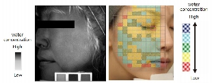
This technique is based on near infrared spectroscopy, which is highly sensitive in detecting water. The distribution of water in the face was able to be visualized with this technique. (Figure 1)
[Spontaneous Raman scattering spectroscopy]
This is a technique that can detect many functional groups that are unique to skin components, such as proteins, lipids, and amino acids, in addition to water. It was used to elucidate the depth profile of components in the skin, such as water and amino acids.
[Non-linear Raman scattering spectroscopy]
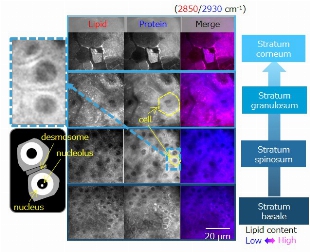
This is a high-speed imaging technique that is about 1,000 times faster than spontaneous Raman scattering spectroscopy. With this technique, the 3D distribution of water, proteins, and lipids in the skin was able to be visualized. (Figure 2)
Figure 1
Distribution of water in the skin as visualized by near infrared spectroscopic imaging
(Right image: Shows the average for multiple individuals; selected from the related research paper (1) described below)
The average distribution of the water in the human facial skin can be analyzed by pseudo-coloring of the water content.
Figure 2
Distribution of components in the skin as visualized by non-linear Raman scattering spectroscopy
(Selected from the related research paper (2) described below)
The state of epidermal metabolism was evaluated at the cellular level by using a non-invasive method to visualize the micro-distribution of skin components such as proteins and lipids.


Related research papers
24 research papers, including the following:
(1) Egawa M et al, Visualization of Water Distribution in the Facial Epidermal Layers of Skin Using High-Sensitivity Near-Infrared (NIR) Imaging. Appl Spectrosc. 2015 Apr;69(4):481-7.
(2) Egawa M et al. Label-free stimulated Raman scattering microscopy visualizes changes in intracellular morphology during human epidermal keratinocyte differentiation. Sci Rep. 2019 Aug 29;9(1):12601.
*The content of the release is correct as of the time of release, but please note that it may in some cases differ from the latest information.
Related News

Jul. 6, 2026
Shiseido Discovers “Ring Collagen®,” a Collagen Structure That Creates Tension to Maintain Facial Shape

May. 25, 2026
Shiseido Wins First Prize at the 2026 China Cosmetics Science and Technology Conference

Apr. 16, 2026
Shiseido Discovers That Age-Related Uneven Arterial Blood Flow Causes Facial Sagging